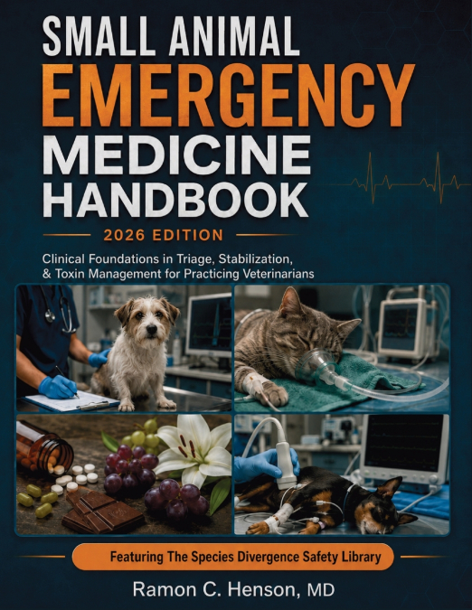
Ramon C. Henson MD
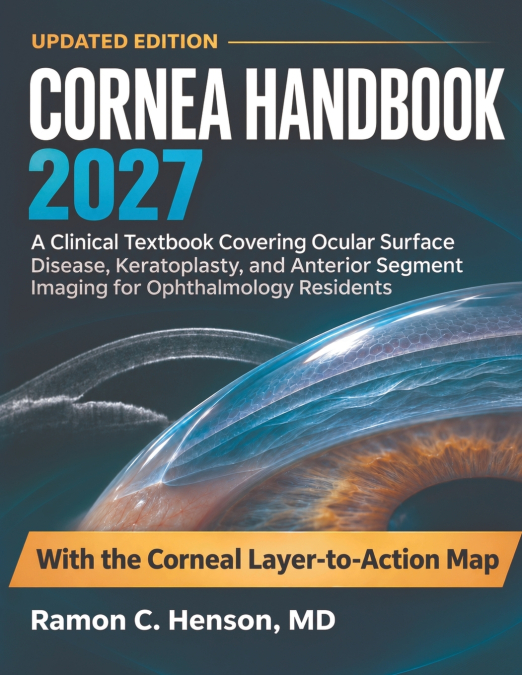
Read the Layer, Recognize the Pattern, Protect the Tissue Built for ophthalmology residents, cornea fellows, and clinicians who must convert subtle anterior-segment findings into timely action, this clinical textbook connects corneal anatomy and wound biology to diagnosis, medical care, optical rehabilitation, and surgery. It follows the full arc of practice: tear-film instability, epithelial failure, microbial and immune keratitis, dystrophies, ectasia, endothelial disease, trauma, refractive complications, keratoplasty, and lifelong graft surveillance. Layer-specific examination, slit-lamp morphology, tomography, anterior-segment OCT, specular microscopy, narrative cases, and structured decision pathways make each finding accountable to mechanism and trajectory. What This Book Puts Into Clinical Practice Localize pathology by tissue layer - distinguish epithelial, stromal, endothelial, interface, and anterior chamber processes before treatment begins Separate sight-threatening infection - integrate ulcer geography, infiltrate depth, host risk, sampling strategy, and early response Interpret shape and thickness imaging - connect topography, tomography, pachymetry, and epithelial maps to ectasia progression Restore the failing surface - manage dry eye phenotypes, neurotrophic disease, persistent defects, exposure, and medication toxicity Choose the appropriate graft - match PK, DALK, DSAEK, or DMEK to diseased tissue, recipient anatomy, and visual potential Recognize postoperative danger early - detect detachment, rejection, infection, pressure injury, suture complications, and graft failure Use the Corneal Layer-to-Action Map - move from visible signal through mechanism and pattern to an explicit action threshold and safety boundary Bring clearer structure to the next corneal finding and make every follow-up test a defensible clinical prediction.